Discovering new details of the human intestinal mesentery

A new discovery regarding the human intestinal mesentery may radically change the fundamentals of anatomy and embryology and related surgical approaches.
The mesenery is an obscure structure in the human body that was largely unknown until recently. Researchers have known about this body part for centuries, but not in the exact details recently uncovered in an observational study with human cadavers.
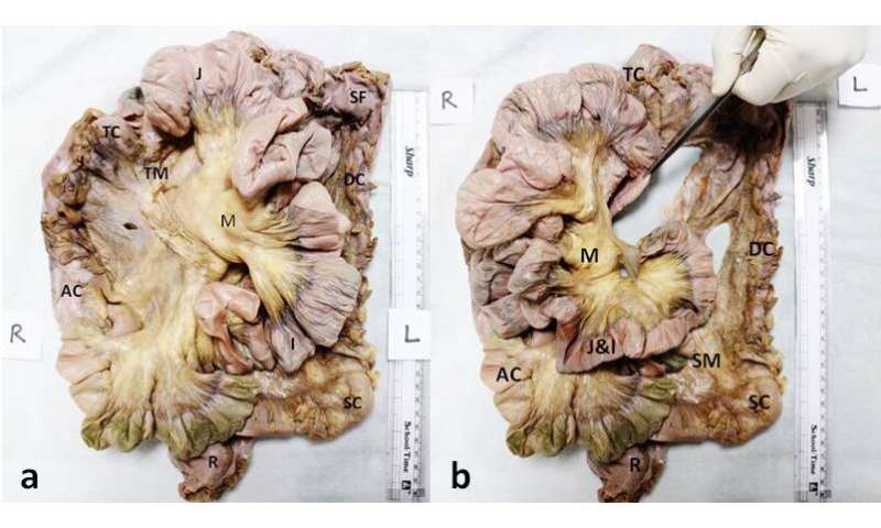
The mesentery is a double fold of a membrane (the peritoneum) that supports the intestine from the posterior abdominal wall carrying vessels and nerves. During early development, the whole of the gut tube that forms the intestine bears mesentery, but it is thought to degenerate for many parts of the intestine as development advances. Mesentery has, until recently, been regarded as a fragmented structure and present only in some parts of the intestine in adult humans. Now, in the new study, we have found a continuous mesentery present all along the intestine (Fig.1).
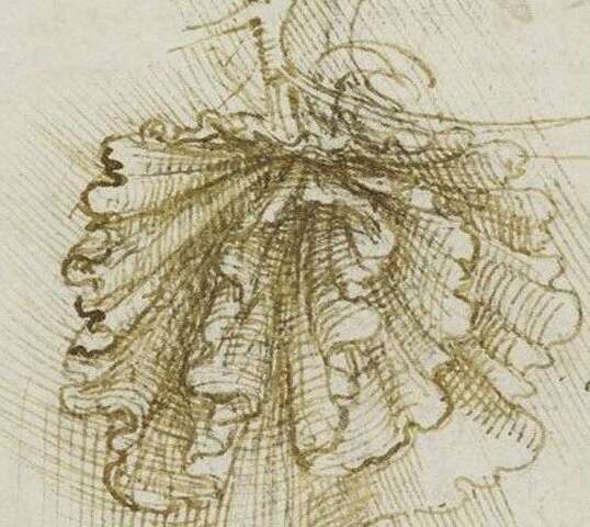
An indication of continuous mesentery can be inferred by a sketch drawn by Leonardo Da Vinci in 1452 and a text description by anatomist Carl Toldt in 1879. Then, in 1885, Sir Frederick Treve gave a series of lectures at the Royal College of Surgeons in London during which he described a fragmented mesentery in adult humans. Since then, this view has been reported in standard anatomy and embryology textbooks. Recently, this model was challenged by Professor Calvin Coffey and colleagues from the University Of Limerick, Ireland, with the suggestion that the mesentery is continuous. They observed intra-operative and radiological evidence that the human mesentery is continuous from the duodenojejunal junction to the rectum.

Professor Coffey's observations inspired a new series of research, as cadaveric proof for the continuous mesentery was still lacking, and how to dissect an intact mesentery was not known. Anatomists' ignorance about the presence of intact mesentery in adults was surprising. We thought that the conventional method used to dissect mesentery, which was inspired by Treves, might be the reason that we were missing certain structural details.
In 2017, we revised the existing method for the mesenteric dissection and reported a continuous mesentery in two cadavers. This was perhaps the first study in cadavers showing an intact mesentery (see video). Surprisingly, we not only confirmed Professor Coffey's intraoperative and radiological observations, but additionally made a completely new finding that the intestinal mesentery continued up to the gastroduodenal junction, which meant that the duodenum contained a mesentery in adults. This was completely new information in human anatomy, which enthralled us, and recalls the sketch by Leonardo Da Vinci showing an intact mesentery for the complete intestine (Fig. 2).
We realized that we had discovered the mesentery as depicted in Da Vinci's sketch. Professor Coffey later joined us in this endeavor, and together, we studied mesentery structures in 18 more cadavers—in fact, we found an intact mesentery in the full length of the intestine in 100% of the cases, which strongly suggests it is present in all individuals.
We have inspired a conceptual change in abdominal anatomy, and have laid the long-held debate to rest by validating that the mesentery is a single structure. The findings of our second observational study have been now published in the journal Frontiers in Surgery.
This research is tantamount to a paradigm shift in the surgical approaches to the intestine. A continuous mesentery provides a wonderful opportunity for the surgeons to follow an avascular anatomical plane around the mesenteric sheath to reach different parts of the intestine, thereby simplifying surgical approaches. Ease of access for the target part of the intestine and a safer route for traversing along the mesenteric plane will not only reduce the time of surgery, but also morbidity rates due to less chance of bleeding and intraoperative complications. An intact mesentery-based surgical approach may be of immense help in limiting the spread of colorectal and other abdominal cancers, and clearing affected lymph nodes.
Another study of the gross anatomy of the mesentery with respect to its length, continuity and attachments is of great significance, since it could provide a better understanding of congenital gastrointestinal anomalies like malrotation and malfixation of the intestine after its rotation during the embryonic development. A continuous mesentery is also likely to reveal the mechanism by which the persistence of the embryological intestinal anomalies lead to obstructions, and sometimes give rise to surgical emergencies in adults.
Being highly vascular, it has also a potential role in the metabolism of the drugs. The mesentery is known to contain immune cells in abundance, a phenomenon said to be implicated in the pathogenesis of inflammatory bowel diseases like Crohn's disease and ulcerative colitis. A detailed description of the extensive anatomy of the human mesentery would contribute to further immunological research of this hitherto ignored body part.
The study has inspired many other conceptual changes in anatomy and surgery textbooks, including the neurovascular distribution of the mesentery, attachment of the mesentery to the abdominal wall, and disposition of the peritoneum around the intestine and other abdominal organs.
This story is part of Science X Dialog, where researchers can report findings from their published research articles. Visit this page for information about ScienceX Dialog and how to participate.
More information: 1. Kumar A, Faiq MA, Krishna H, Kishan V, Raj GV, Coffey JC and Jacob TG (2020) Development of a Novel Technique to Dissect the Mesentery That Preserves Mesenteric Continuity and Enables Characterization of the ex vivo Mesentery. Front. Surg. 6:80. DOI: 10.3389/fsurg.2019.00080
2. Coffey JC, O'Leary DP. The mesentery: structure, function, and role in disease. Lancet Gastroenterol Hepatol. 2016;1(3):238–247. DOI: 10.1016/S2468-1253(16)30026-7
3. Kumar A, Kishan V, Jacob TG, Kant K, Faiq MA. Evidence of continuity of mesentery from duodenum to rectum from human cadaveric dissection—a video vignette. Colorectal Dis. 2017;19(12):1119–1120. DOI: 10.1111/codi.13917
4. Kumar A, Ghosh SK, Faiq MA, Deshmukh VR, Kumari C, Pareek V. A brief review of recent discoveries in human anatomy. QJM. 2019;112(8):567–573. DOI: 10.1093/qjmed/hcy241
Bio:
Ashutosh Kumar is an Assitant Professor in the Department of Anatomy, All India Institute of Medical Sciences, Patna, India. Muneeb A. Faiq is a postdoctoral researcher in Langone Health Centre, New York University (NYU), New York, USA.