The growth and differentiation potential of dental pulp cells in demineralized dentin tubules

Scientists associated with regenerative therapy have attempted to regenerate pulp-dentin complex due to the damage caused by infection, trauma or developmental anomaly of permanent teeth with necrotic pulp. Therefore, with the concept of tissue engineering of stem cells, scaffolds and signaling molecules, true pulp regeneration seem to be an achievable goal.
Therefore, the group of researchers from Graduate School of Tokyo Dental College, Ziauddin University, Hayatabad Medical Complex, and King Saud University performed a study on the dental pulp cells.
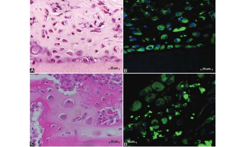
In their study, published in BJBMS, the researchers attempted to regenerate a biocompatible osteodentin-like hard tissue matrix in order to replace gutta-percha which is used in the conventional root canal treatment. In this study, they confirmed, using immunohistochemical analysis, that dentin-like hard tissue was produced that was composed of dentin forming cells including odontoblasts, and bone-producing cells osteoblasts.
The researchers transplanted dental pulp cells embedded in the collagen-type gel and loaded into demineralized dentin tubules. Thirty-two Sprague-Dawley rats were used in this experiment, while six green fluorescent protein-transgenic rats were used as donor for the collection of pulp cells.
After three weeks of the experiment, the researchers found osteodentin-like hard tissue inside the demineralized dentin tubules. Osteodentin-like hard tissue matrix was immunohistochemically analyzed with the bone and dentin antibodies like alkaline phosphatase, bone sialoprotein, osteopontin, nestin, and dentin sialoprotein.
Generally, dental pulp stem cells with scaffold and growth factors are used in regenerative endodontic therapy. In this study however, instead of stem cells, the researchers used cultured or expanded pulp cells up to third passage and growth factor of bone morphogenetic protein expressed within the surface of the root canal with EDTA.
With further experimental research studies, this study model can be used to develop ex vivo graft material of pulp cells embedded in collagen type-1 gel and chelation of the inner surface of the conventional root canal. Subsequently, this graft material can be loaded into sterilized empty root canal using injection method which can produce osteodentin-like hard tissue to replace gutta-percha in root canal treatments.
More information:
Zeb Khan S, Mirza S, Karim S, Inoue T, Bin-Shuwaish MS, Al Deeb L, Al Ahdal K, Al-Hamdan RS, Maawadh AM, Vohra F, Abduljabbar T. Immunohistochemical study of dental pulp cells with 3D collagen type I gel in demineralized dentin tubules in vivo. Bosn J of Basic Med Sci. 2020
Provided by Association of Basic Medical Sciences of FBIH