Kazan Federal University scientists studying extracellular matrix and microglial cells in the injured spinal cord to dev

Scientists from the Institute of Fundamental Medicine and Biology of Kazan Federal University in collaboration with Russian and British colleagues, characterized the state of the extracellular matrix, as well as auxiliary cells that ensure the functioning of the spinal cord after its traumatic injury. New data will be relevant in the development of more effective methods for restoring lost motor functions after neurotrauma. The results of this research, supported by grants from the Russian Science Foundation and the Russian Foundation for Basic Research, were published in highly rated international scientific journals Frontiers in Cellular Neuroscience and Frontiers in Molecular Neuroscience.
Two independent projects implemented as parts of the work of the one scientific group "Molecular and cellular mechanisms of neuroregeneration", headed by doctor of medical sciences, Leading Researcher of the Laboratory of Gene and Cell Technologies Yana Mukhamedshina, were aimed at understanding the role of nerve cells in maintaining the extracellular matrix* of the spinal cord after its damage, as well as the characteristics of a special type of cells—microglial cells in the injured spinal cord, depending on the severity and terms of the injury.
* The extracellular matrix is a multicomponent substance in which all cells of the body are immersed. In the last decade, interest in the extracellular matrix has increased significantly. This is due to the establishment of its role in aging, regeneration, cancer therapy, and the treatment of certain hereditary diseases.
"The gist of the two projects is that the cells in the central nervous system, depending on the signals around them, can play both a positive and a negative role in restoring motor function after a spinal cord injury. Until now, it remained unclear how the behavior and phenotype of various nerve cells change when the spinal cord is injured, and whether these indicators depend on the severity of the injury, and how far the cells are located from the epicenter of the injury," explains Dr. Mukhamedshina.
Spinal cord injury leads to a pronounced restructuring of the nervous tissue with the formation of a scar in the damaged area, which blocks the growth of axons and the restoration of nerve connections. Changes in the state of cells and the composition of the extracellular matrix in the nervous tissue remote from the epicenter of damage in different periods after injury have not been well studied. "That is why we studied the cellular and molecular composition of this area on the model of contusion spinal cord injury of the rat in the ventral horns, where the motor neurons responsible for muscle innervation and limb movement are located, by various methods of analysis," says co-author, 4th year Ph.D. student, Junior Researcher of the Laboratory of Gene and Cell Technologies Kabdesh Ilyas. "Our data draw attention to the different types of cells that are involved in the restructuring of the extracellular matrix of the nervous tissue both in the damaged area and in a more intact tissue at a distance from the neurotrauma."
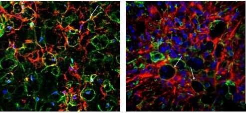
Lately, more scientists have been confirming the key role plated by microglial cells in the pathological processes emerging from a spinal cord injury. Depending on its surroundings, microglia can assume either a toxic or a protective phenotype. Additionally, it has been shown that both phenotypes are mutually transferrable. "So far there have been no data about the modulations of microglial cells after neurotrauma and the characteristics of such changes under different severity," adds another co-author, 4th year Ph.D. student, Junior Researcher of the Laboratory of Gene and Cell Technologies Elvira Akhmetzyanova. "In our study, we tried to reveal such mechanisms and for a first time described annular-shaped microglial cells, the morphology of which is shaped by the spatial orientation of cell outgrowths forming round or oval micro-territories with disintegrating myelin fibers inside them. This type of cell morphology was found only in the injured spinal cord and mainly in the white matter."
As the authors point, the results are important for further studies of pathophysiology of spinal cord injury and pathological reactions of the nervous system after local damage. The practical significance is that the research shows potential cellular and molecular targets in areas distant from the injury, which is important for an extended growth of axons and a more comprehensive recovery of motor functions.
More information:
Cellular and Molecular Gradients in the Ventral Horns With Increasing Distance From the Injury Site After Spinal Cord Contusion
www.frontiersin.org/articles/1 … cel.2022.817752/full
Increasing Severity of Spinal Cord Injury Results in Microglia/Macrophages With Annular-Shaped Morphology and No Change in Expression of CD40 and Tumor Growth Factor-β During the Chronic Post-injury Stage
www.frontiersin.org/articles/1 … mol.2021.802558/full
Provided by Kazan Federal University