Read the invisible pores in CT images

Porous materials are ubiquitous—from ceramics to soils and rocks to human bone and various components, that are everywhere in daily life and are critical to virtually medical and all industrial construction and energy processes. Many construction materials are porous, such as typical concrete and cement. In recent years, with increasing interest in the lunar exploration and base construction, building materials manufactured through sintering of the in situ resources (i.e., lunar regolith) and characterization on the sintered porous materials have attracted more and more attention. Most porous materials are composed of matrix and pores at microscale or nanoscale which are invisible to human eyes.
Most people heard of CT (Computed Tomography) scan, when they are getting the physical examination in the hospital. X-ray CT has been widely used to analyze porous materials for its major advantage of quantitative measurement of pore structures even at nanoscopic scale. The Korea Institute of Civil Engineering and Building Technology (KICT, President Kim Byung-Suk) owns a high-tech multi-tube industrial CT scanner, which has been successfully applied in various research fields and provided services for many research entities and enterprise in domestic and overseas. Over years' experience in running CT scanning and image processing lay a sound foundation for developing new analysis method of material characterization.
The KICT announced that the research team led by Dr. Hyusoung Shin has developed a new method, named the statistical phase fraction (SPF) method, to estimate porosity and to evaluate homogeneity of porous materials dominated by sub-resolution pores via CT image analysis.
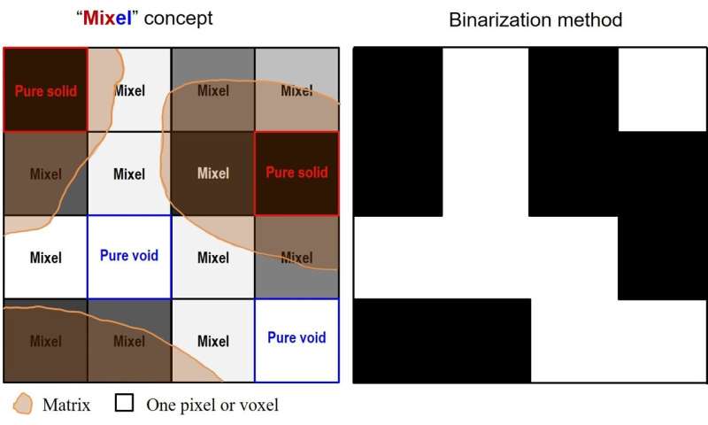
A traditional and the simplest segmentation technique is the binarization method, where a threshold pixel intensity value is selected to divide the image into two portions and pores are usually grouped into the portion that has pixel values smaller than the threshold. However this approach can only deal with pore size that is great than or equal to 1 pixel. The research team introduces a new term of "Mixel" (Fig. 1), which represents a pixel or a voxel consisting of two or more phases. This method employs Gaussian function fitting on the CT histogram and the CT number for each single phase (e.g., air, water, pure solid) that was included in the sample. The total porosity for any given bulk volume with irregular shape can be estimated.

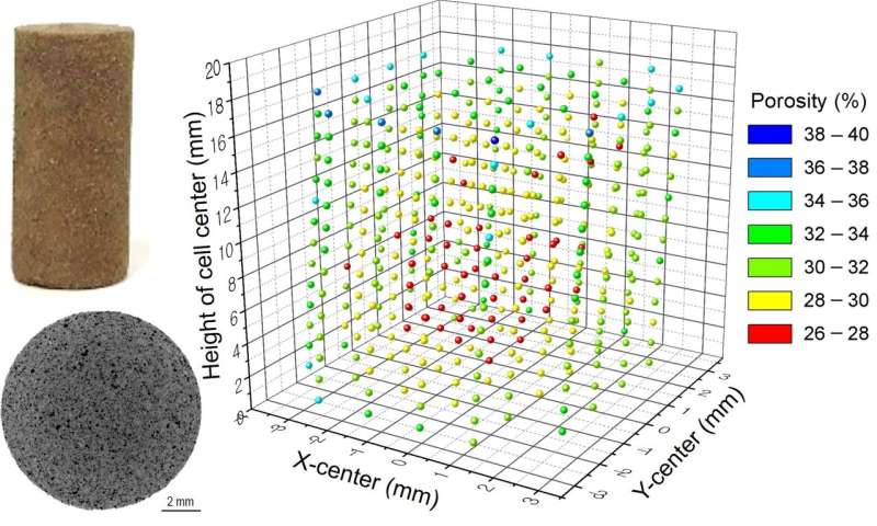
Based on a lot of trial and error crossing many years, this method has been successfully applied in estimation of porosity and evaluation of homogeneity of sintered lunar regolith simulant, which is expected to be a candidate construction material for future moon construction (Fig. 2). Dr. Li Zhuang from KICT commented that "The SPF method has a big advantage over existent methods that it can estimate local porosity for any arbitrary part of a given sample without destroying the sample. This is extremely useful for evaluation of sample homogeneity." The research team saw great potential in the newly developed SPF method for its applications to other porous fine-grained materials, such as bentonite and engineered cement used for construction materials of nuclear waste disposal facilities.
Provided by National Research Council of Science & Technology