Using infrared cameras to detect early stage breast cancer

Cancer is one of the most debilitating diseases in the world, affecting hundreds of thousands of people every year. In the United States, cancer is the second-highest leading cause of death. Further, breast cancer affects one in eight women within the United States every year. While the causes of breast cancer are still unknown, options in screening and treatment have improved greatly over time. The earlier breast cancer is detected, the faster it can be cured.
Currently, there are several methods used worldwide. Mammography is the most widely used method, using X-rays and compressing the breasts to get a better picture. Normally, a tumor appears as a solid white mass, however, in denser breast tissue, detecting these masses becomes very difficult. Additionally, the discomfort and radiation associated with mammography often discourages women from annual screening. Ultrasound provides a non-invasive, inexpensive option that can distinguish between a malignant, palpable tumor and a benign, fluid-filled cyst. Unfortunately, it is highly user-dependent and can often result in misdiagnosis. Finally, magnetic resonance imaging (MRI) is primarily used for high-risk screening (known or suspected mutation in a breast cancer causing gene or lifetime risk > 20%) and further diagnosis prior to surgical resection. MRIs are not generally used for everyday screening due to high cost and false positives. Another factor that influences many screening methods is the tissue density of the breast. Over 40% of the population has denser breast tissue, and heterogeneously and extremely dense breast tissue can often mask potential tumors on mammography.
The Thermal Analysis Lab (TAMFL) at the Rochester Institute of Technology in Rochester, New York, has collaborated with a team of clinicians from the Rochester General Hospital (RGH) to develop a noninvasive breast cancer screening method. The team consists of Dr. Satish Kandlikar (RIT—professor), Dr. Pradyumna Phatak (RGH—chief of medicine), Dr. Lori Medeiros (RGH—medical director of Breast Center and breast surgeon), Dr. Donnette Dabydeen (RGH—consulting radiologist), the RGH clinical research team (Tia Albro, Kayla Malchoff, Megan Littleton, and Kellie Sherron) and several graduate students at RIT (Jose Luis Gonzalez-Hernandez, Carlos Gutierrez and myself). The TAMFL-RGH team is working on a method called steady-state infrared imaging (IRI).

This method is by no means new to breast imaging and has actually been used since the 1950s. IRI, also known as thermography, is particularly attractive due to lack of ionizing radiation, patient comfort (the technology is touchless and does not require breast compression), and portability. Although thermography was approved as an adjunctive technique by the Food and Drug Administration (FDA) in 1982, it has fallen out of favor due to poor accuracy and nonstandardized clinical protocol.
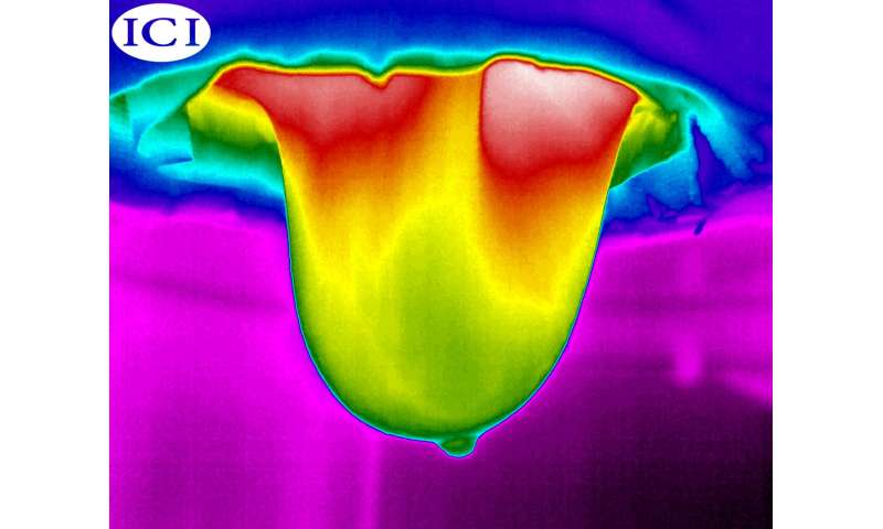
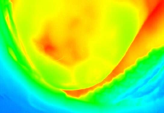
However, the collaborative study we have undertaken is focusing on optimizing the technology from both an engineering and medical standpoint. Infrared imaging has the unique ability to capture thermal activity in breast tissue resulting from a tumor. The growth of a tumor is associated with inflammatory properties and an increase in blood and oxygen, which causes a larger temperature change between the growing tumor and the surrounding breast tissue. Thermal activity such as cellular metabolism, increased vasculature and increased blood perfusion create areas of increased temperature. The generated heat appears at the breast surface and the temperature changes are detected with an infrared camera. The new project has combined heat transfer fundamentals, tumor pathology and engineering design to create a method that is scientifically based and validated by current gold standard screening modalities.
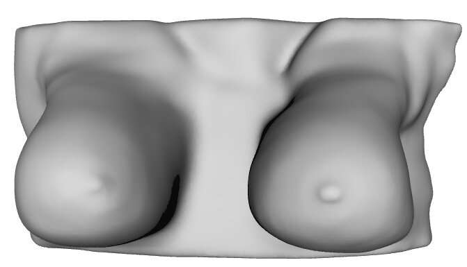
The first phase of the TAMFL-RGH study used patients with known biopsy-proven breast cancer. The ultimate goal was to test the method and validate using a well-known, trusted modality. Using patients with known breast cancer, we were able to screen after a pre-surgery MRI and before surgical resection. When women are screened for breast cancer using MRI, they are screened in the prone position (on their stomach) to allow the breasts to hang freely, uninhibited by gravitational deformation. This same position was used for IRI for a more accurate comparison. To analyze the images, a patient-specific computer model of the breast is created. Using this model and the collected infrared images, we are able to pinpoint the exact position and size of the tumor. So far, we have tested this in seven patients with biopsy-proven breast cancer. This method has identified the size and position of the tumor in the cancerous breast and has accurately identified "no cancer" in the healthy breast. This work has been published in several journals.
The second phase of the study is to enroll more patients, enroll patients who do not have breast cancer and optimize the technology for more rapid diagnosis. This phase the study will allow us to determine the efficacy of the technology and the ability to differentiate between a malignant tumor and healthy tissue. Many of the patients we recruit may have a benign mass or a false positive. Imaging these patients will further establish IRI as a potential adjunctive technique for breast cancer screening. Unfortunately, due to the coronavirus pandemic, screening and recruitment has been temporarily suspended. However, the results from the first phase of the study are highly encouraging and will hopefully lead to a large-scale clinical study.

I am an engineering Ph.D. student at the Rochester Institute of Technology. When I finished my bachelor's degree, I promised to use my degree and my skills to help other people. Working in the hospital with breast cancer patients has been incredibly humbling. I have had family members with cancer and have felt the impact of a loved one struggling against a terrible disease. But working on this study has shown me another side of cancer, a side filled with virtue and strength. Many of the women I have screened are in a whirlwind, about to get a tumor removed or their entire breast removed. It has been an incredibly humbling experience working with these patients, all of whom are volunteers. I feel strongly that the goal of engineering is to contribute to the improvement of people's lives wherever they exist on this planet. While this technology is still in the early stages, it has immense potential. In the words of one of the powerful women I have screened, "This may not help me, but it could help my granddaughter."
This story is part of Science X Dialog, where researchers can report findings from their published research articles. Visit this page for information about ScienceX Dialog and how to participate.
More information: [1] A. N. Recinella, J.-L. Gonzalez-Hernandez, S. G. Kandlikar, D. Dabydeen, L. Medeiros, and P. Phatak, "Clinical Infrared Imaging in the Prone Position for Breast Cancer Screening—Initial Screening and Digital Model Validation," ASME J of Medical Diagnostics, vol. 3, no. 1, Feb. 2020, DOI: 10.1115/1.4045319.
[2] J.-L. Gonzalez-Hernandez, A. N. Recinella, S. G. Kandlikar, D. Dabydeen, L. Medeiros, and P. Phatak, "An inverse heat transfer approach for patient-specific breast cancer detection and tumor localization using surface thermal images in the prone position," Infrared Physics & Technology, vol. 105, p. 103202, Mar. 2020, DOI: 10.1016/j.infrared.2020.103202.
[3] J.-L. Gonzalez-Hernandez, S. G. Kandlikar, D. Dabydeen, L. Medeiros, and P. Phatak, "Generation and Thermal Simulation of a Digital Model of the Female Breast in Prone Position," Journal of Engineering and Science in Medical Diagnostics and Therapy, vol. 1, no. 4, p. 041006, Nov. 2018, DOI: 10.1115/1.4041421.
Bio:
Alyssa Owens is a PhD student at the Rochester Institute of Technology in Rochester, NY. She has a background in mechanical engineering and experience with heat transfer in fluids research. For her PhD, she has been fortunate to change fields and focus on breast cancer screening. As a member of the IRI Breast Cancer Team, she has been responsible for collaborating with doctors and screening patients with infrared imaging or thermography. Alyssa is hoping to eventually continue this research with the goal of creating a company or product.